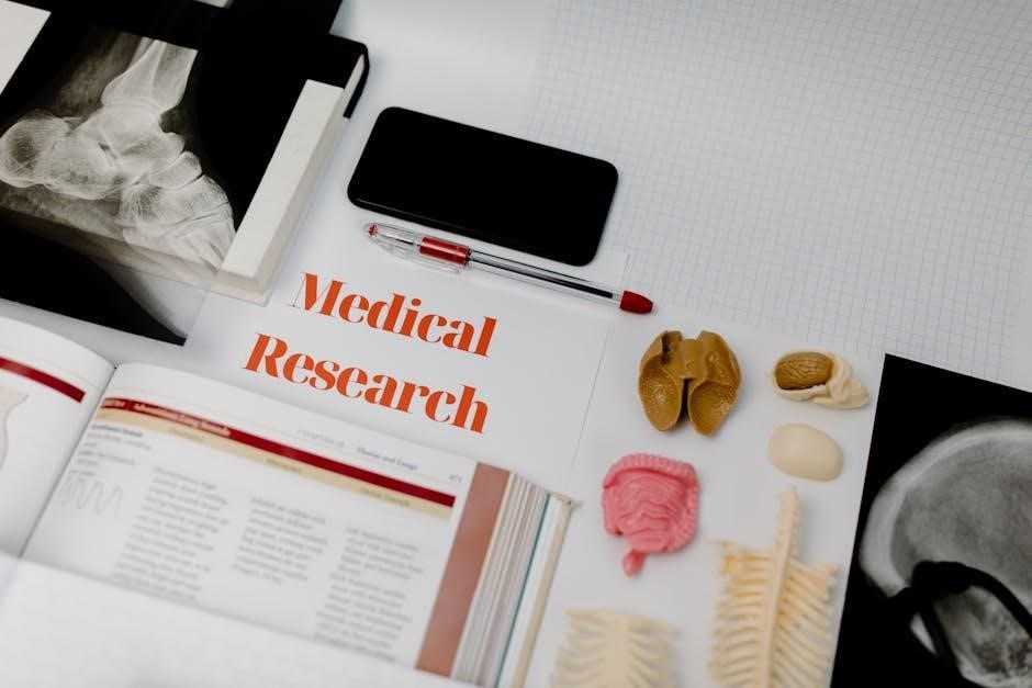
Welcome to the Anatomy & Physiology Lab Manual, designed for a two-semester course. This comprehensive guide blends hands-on activities with digital simulations, providing a dynamic learning experience.
1.1 Purpose and Scope of the Lab Manual
This manual is designed for a two-semester Anatomy & Physiology course, providing hands-on activities, digital simulations, and customizable content. Its purpose is to enhance understanding of the human body’s structure and function, offering practical exercises and tools for healthcare students to master anatomical concepts effectively.
1.2 Importance of Lab Work in Anatomy & Physiology
Lab work enhances understanding of human anatomy and physiology by providing hands-on experience. It allows students to visualize structures, perform experiments, and observe physiological processes, reinforcing theoretical knowledge and improving retention through practical application and interactive learning experiences.
1.3 How to Use the Lab Manual Effectively
To maximize the lab manual’s benefits, follow structured exercises, review safety protocols, and utilize tear-out worksheets. Engage with digital tools like eLab modules and interactive 3D models for enhanced learning. Familiarize yourself with the manual’s layout and customize activities to align with your course goals for optimal outcomes.
Lab Safety and Preparation
Lab safety is crucial for protecting students and equipment. Always wear PPE, follow protocols, and handle tools carefully. Prepare by reviewing instructions and organizing materials to ensure efficient experimentation.
2.1 Safety Protocols in the Lab
Adhering to safety protocols is essential in the anatomy and physiology lab. Students must wear protective gear like gloves and goggles. Proper handling of equipment and chemicals ensures a safe environment. Familiarize yourself with emergency procedures and maintain a clean workspace to prevent accidents and infections.
2.2 Handling Lab Equipment and Tools
Proper handling of lab equipment is crucial for safety and accuracy. Microscopes, dissecting tools, and other instruments require careful use and maintenance. Always follow manufacturer guidelines and clean equipment after use to ensure functionality and prevent contamination. Familiarize yourself with each tool’s purpose before starting experiments.
2.3 Preparing for Lab Experiments
Thorough preparation is essential for successful lab experiments. Review lab materials, safety guidelines, and objectives beforehand. Gather all required supplies and ensure equipment is in proper working condition. Familiarize yourself with procedures and wear appropriate lab attire. Understanding pre-lab steps enhances efficiency, safety, and learning outcomes in anatomy and physiology experiments.
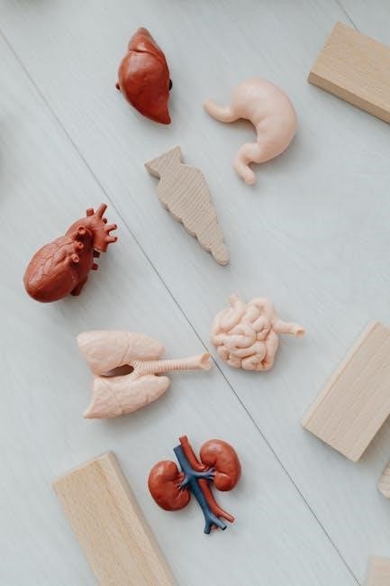
Histology and Microscopy
Histology involves the study of tissue structure under a microscope. This section introduces microscopy techniques, tissue preparation, and examination methods to understand cellular and tissue organization in anatomy.
Histology is the study of tissue structure and organization. This section introduces students to the fundamentals of histology, including tissue types, preparation methods, and microscopic examination techniques. Understanding histology is crucial for analyzing anatomical structures and their physiological functions.
3.2 Setting Up and Using a Microscope
Mastering microscope setup is essential for histological observations. Start by adjusting the lenses and focusing the stage. Properly align the light source and use the coarse and fine adjustment knobs for clarity. Practice slide preparation and staining techniques to ensure high-quality tissue visualization during lab exercises.
3.3 Preparing Tissue Samples for Examination
Preparing tissue samples involves sectioning, staining, and mounting to ensure clear microscopic observation. Follow proper histological techniques to maintain tissue integrity. Safety protocols must be adhered to during handling and processing. Accurate preparation enhances the visibility of cellular structures, aiding in detailed anatomical and physiological analysis.
The Integumentary System
This section explores the skin and its appendages, focusing on their structure, function, and role in protecting the body and regulating vital physiological processes.
4.1 Structure and Function of the Skin
The skin, the body’s largest organ, consists of the epidermis, dermis, and subcutaneous layers. It protects against pathogens, regulates temperature, and aids in sensation, immune response, and vitamin D synthesis through specialized cells like keratinocytes and melanocytes.
4.2 Identifying Skin Layers and Appendages
The skin comprises the epidermis, dermis, and subcutaneous layers. Appendages include hair follicles, sweat glands, and nails. Lab exercises involve identifying these structures, understanding their functions, and exploring their roles in protection, sensation, and bodily regulation through microscopy and dissection activities.
4.3 Lab Exercises on Skin and Surface Anatomy
Lab exercises focus on hands-on exploration of skin layers and surface anatomy. Students identify epidermal and dermal features, examine appendages like hair and nails, and use microscopy to visualize cellular structures. These activities enhance understanding of the skin’s protective and sensory functions through practical observation and analysis.
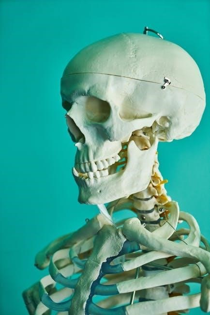
The Skeletal System
Explore the skeletal system, focusing on bones and joints. Labs include identifying axial and appendicular skeleton components, enhancing understanding of structural and functional roles through hands-on activities.
5.1 Overview of Bones and Joints
Bones are rigid, calcified structures providing support and protection. Joints are points where bones connect, enabling movement. This section introduces bone classification, joint types, and their functions. Hands-on activities, such as bone identification, enhance understanding of skeletal structure and joint mobility in the human body. Explore the composition of bones and the role of joints in facilitating movement.
5.2 Axial and Appendicular Skeleton
The axial skeleton includes the skull, vertebral column, and ribcage, forming the body’s central framework. The appendicular skeleton comprises the upper and lower limbs and pelvis, enabling movement. Labs focus on identifying bone structures and understanding their roles in providing stability and facilitating motion.
5.3 Lab Activities for Bone Identification
Lab activities focus on identifying and analyzing human bones, such as the sacrum, humerus, and femur. Students learn to recognize key features, like foramen and epicondyles, to distinguish bones. Hands-on exercises with skeletal models and digital tools enhance understanding of bone structure and function in the skeletal system.
The Muscular System
This section explores the structure and function of muscles, including skeletal, smooth, and cardiac types. Lab activities involve identifying major muscle groups and understanding their roles in movement and body support.
6.1 Types of Muscles and Their Functions
The muscular system comprises skeletal, smooth, and cardiac muscles. Skeletal muscles enable voluntary movement and posture support. Smooth muscles perform involuntary actions like digestion. Cardiac muscle powers the heart’s rhythmic contractions, ensuring blood circulation. Each type has unique structures and roles tailored to their specific physiological functions.
6.2 Muscle Actions and Movements
Muscles produce movement through contraction, enabling actions like flexion (bending) and extension (straightening). Abduction moves limbs away from the midline, while adduction brings them closer. These movements are essential for locomotion, posture, and daily activities, and are explored through lab exercises to enhance understanding of muscular mechanics and function.
6.3 Identifying Major Muscle Groups
The lab manual guides students in identifying major muscle groups, such as the pectoralis major, quadriceps, and gastrocnemius. Through dissection, 3D models, or digital simulations, students learn to locate and differentiate muscles, enhancing their understanding of muscle structure, function, and role in movement and stability.
The Nervous System and Senses
This section explores the structure and function of the nervous system, including nerve tissues, reflexes, and sensory organs, enhancing understanding through practical lab exercises.
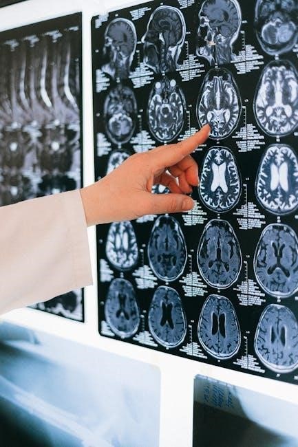
7.1 Structure and Function of the Nervous System
This section delves into the intricate organization of the nervous system, detailing its components, such as neurons, glial cells, and synapses, while explaining how these elements collaborate to enable sensory perception, motor responses, and complex cognitive functions through interactive lab activities and detailed illustrations.
7.2 Identifying Nerve Tissues and Reflexes
Explore nerve tissues and reflexes through interactive lab exercises. Activities include histological preparations to examine nerve structures and reflex testing to observe motor responses. These hands-on experiences, combined with virtual simulations, offer a comprehensive understanding of neural mechanisms, reflex pathways, and their physiological significance.
7.3 Lab Exercises on Sensory Organs
Lab exercises focus on exploring sensory organs through histological slides and interactive simulations. Students identify structures like the eye and ear, analyze sensory pathways, and conduct reflex tests. These activities enhance understanding of sensory mechanisms and their physiological roles in maintaining homeostasis and nervous system function.
Dissection Exercises
Dissection exercises provide hands-on experience with anatomical structures, emphasizing safety and proper techniques. These labs offer a detailed exploration of tissues and organs, enhancing practical understanding.
8.1 Preparing for Dissection
Preparing for dissection involves gathering necessary tools, reviewing procedures, and ensuring proper equipment handling. Safety attire, including gloves and goggles, is essential. The workspace should be clean and organized, with all materials within reach. Pre-lab review of anatomical structures enhances focus and efficiency during the exercise.
8.2 Conducting Dissections Safely
Conducting dissections safely requires careful handling of tools like scalpels and forceps. Always wear gloves and goggles to protect against exposure. Follow established protocols for specimen handling and maintain focus to avoid accidents. Ensure all sharps are managed securely, and never touch the dissecting tools’ edges carelessly.
8.3 Post-Dissection Procedures and Cleanup
After dissection, properly dispose of specimens and biological waste. Clean and disinfect tools, workstations, and equipment. Store instruments in designated areas and sanitize gloves and goggles. Ensure all materials are labeled and securely stored. Follow lab protocols for waste disposal to maintain a safe and organized environment.
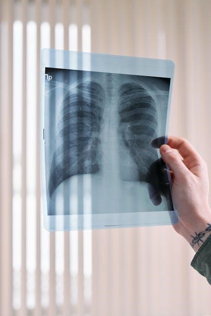
Physiology Experiments
This chapter explores human physiological processes through hands-on experiments. Students measure blood pressure, heart rate, and respiratory functions, gaining practical insights into bodily functions and mechanisms.
9.1 Measuring Blood Pressure and Heart Rate
Students learn to measure blood pressure using a sphygmomanometer and stethoscope, and heart rate through palpation. These exercises emphasize accurate data collection and understanding physiological norms, preparing for real-world healthcare applications.
9.2 Respiratory System Experiments
Experiments focus on measuring lung capacities and gas exchange. Students use spirometry to assess tidal volume, vital capacity, and forced expiratory volume. These activities enhance understanding of breathing mechanisms and the physiological processes essential for oxygen delivery and carbon dioxide removal.
9.3 Cellular Physiology and Histology
Experiments explore cellular structures and functions, such as staining techniques to observe tissue samples under microscopes. Activities emphasize histological processes, linking cellular anatomy to physiological mechanisms. Practical exercises help students understand how cells contribute to overall bodily functions and maintain homeostasis.
Digital Tools and Simulations
Digital tools and simulations enhance learning through virtual dissections, physiology experiments, and interactive 3D models. These resources provide immersive experiences, enabling students to visualize complex anatomical structures and physiological processes in detail.
10.1 Using Virtual Dissection Software
Virtual dissection software provides detailed 3D models of human anatomy, allowing precise exploration of structures. Interactive tools enable students to manipulate specimens, enhancing traditional dissection experiences. This resource fosters deeper understanding and engagement, especially for complex anatomical systems, making it an invaluable supplement to hands-on lab work.
10.2 Physiology Lab Simulations
Physiology lab simulations, such as PhysioEx 10.0, offer interactive exercises that replicate real-world experiments. These simulations allow students to explore complex physiological processes, like blood pressure regulation and nerve impulses, in a controlled digital environment. They enhance learning by providing visual and data-driven insights, complementing traditional lab activities effectively.
10.3 Interactive 3D Models for Anatomy
Interactive 3D models provide detailed visualizations of anatomical structures, enabling students to explore complex systems. These models allow rotation, zooming, and layering to examine organs, tissues, and their spatial relationships. They enhance learning by making abstract concepts tangible and fostering a deeper understanding of human anatomy through interactive exploration.
Effective Study Habits and Test Preparation
Develop a structured study routine, focusing on active learning techniques like flashcards and practice quizzes. Regularly review lab materials and use visual aids to reinforce anatomical concepts, ensuring better retention and exam readiness.
11.1 Strategies for Memorizing Anatomical Structures
To effectively memorize anatomical structures, use active learning techniques such as creating detailed flashcards, grouping terms by function or location, and engaging in regular spaced repetition. Practice labeling diagrams and utilize mnemonics to associate complex terms with memorable phrases or images. Additionally, incorporate digital tools like anatomy apps and interactive 3D models to enhance retention and engagement.
11.2 Reviewing Lab Material for Exams
Reviewing lab material for exams involves systematically revisiting lab reports, focusing on key concepts and experiments. Utilize practice questions and digital tools, such as simulations and 3D models, to reinforce understanding. Prioritize active learning over passive memorization for better retention. Teach concepts to others to deepen your grasp.
11.3 Tips for Success in the Anatomy & Physiology Lab
Stay organized by keeping detailed lab notes and reviewing them regularly. Engage actively in experiments and ask questions to clarify concepts. Use digital tools like simulations and 3D models to reinforce learning. Practice identifying structures and functions regularly to build confidence and mastery over time.